

関連記事
Ultrasonic Gene Fragmenter: A Practical Guide to Controlled DNA Shearing
2026-09-16Making DNA shorter is not the only step that must be taken to prepare nucleic acids for sequencing. The size of the fragments should be within a useful range, suitable for the reactions downstream and have limited variation between samples.
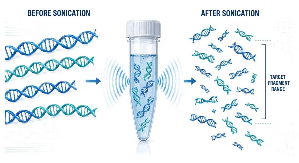
The principle of how an ultrasonic gene fragmenter (more properly called an ultrasonic DNA fragmenter or focused ultrasonicator) works is to break long DNA molecules into shorter fragments, using acoustic energy to do so. With the control of both energy delivery and time and temperature, the process can enable reproducible NGS and chromatin workflows without introducing any sequence specific cleavage.
何 is an Ultrasonic Gene Fragmenter?
An ultrasonic gene fragmenter is one type of instrument used in a laboratory to convey the ultrasonic energy to the biological specimen. Pressure oscillations and cavitation-related mechanical forces shear DNA into smaller fragments.
The instrument can't detect or remove particular genes. The breakage of DNA by ultrasound is generally random, not like that achieved by restriction enzymes or programmable nucleases. Often, the aim is a certain distribution of fragments, instead of a specific nucleotide sequence.
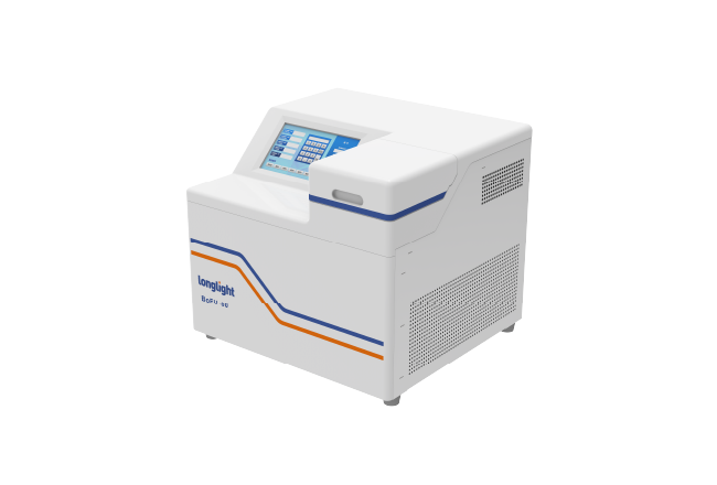
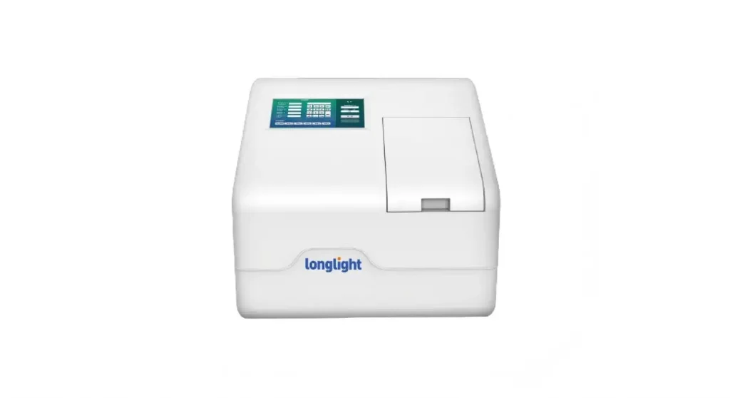
ザ BoFU-100 focused ultrasonicator is specifically optimized for DNA, RNA and chromatin shearing, genome fragmentation and disruption of biological samples by Longlight.
How does Focused Ultrasonication work?
Probe sonicators deliver energy through a probe immersed directly in the sample. Performance can vary with immersion depth, probe condition and operator technique, while direct contact also introduces cleaning and cross-contamination concerns.
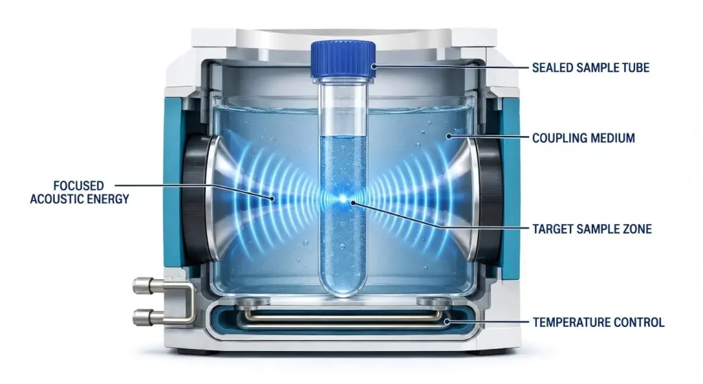
A focused ultrasonicator is a device that focuses the sound energy through a coupling medium to a closed sample tube. Sample is subjected to controlled energy and there is no direct contact between the acoustic source of the instrument and the biological sample.
Processing parameters, such as treatment time, acoustic intensity, duty cycle and temperature, are used to define processing conditions. These variables affect the final fragment distribution and so should be optimized as a whole method and not adjusted alone without verification.
なぜでしょうか 温度 Control is important?
Ultrasonic treatment involves the creation of heat. Excessive sample temperature increases may lead to a decrease in nucleic-acid quality and less predictable final fragment distribution.
An integrated cooling system helps to maintain a more stable processing environment. This is particularly valuable working with low input sample, sensitive chromatin and for multiple sequential runs.
Cooling does not obviate the need for method control. Even with active cooling, sample volume, tube type, starting temperature and processing time should remain consistent because they influence acoustic energy transfer.
What are some of the Applications of Ultrasonic Fragmentation?
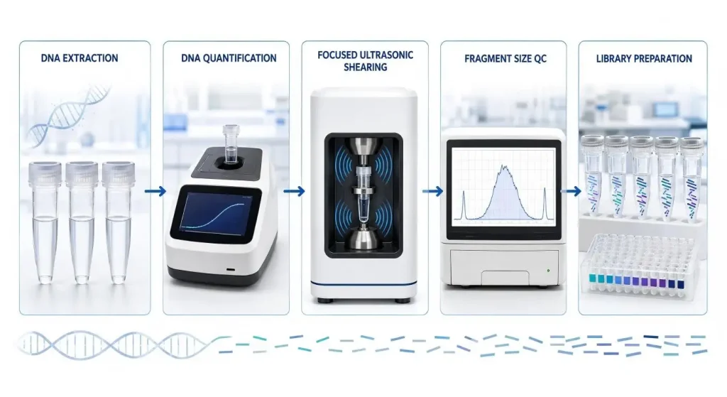
One of the major applications is NGS library preparation. End repair, adapter ligation, amplification or other steps for building the library are performed on a fragmented genomic DNA. The size range desired will vary depending on the particular sequencing platform and protocol used for the library.
Sonication is also required for shearing the cross-linked chromatin in chromatin immunoprecipitation processes. The intention is to generate fragments, which can be immunoprecipitated, that contain relevant DNA-protein interactions.
Other applications may be for RNA fragmentation, FFPE sample processing, for cell and tissue disruption and extraction workflows. Longlight's genomics application solutions include NGS-instruments and lab products.
What Factors Control the ultimate Fragment Size?
Fragment size is dependent on the acoustic energy, treatment time, volume of sample, nucleic-acid concentration, and tube geometry and temperature.
Increasing acoustic energy or treatment time does not necessarily produce a proportional decrease in fragment size. As DNA becomes shorter, fragmentation kinetics change, and excessive processing may reduce recovery or compromise sample quality.
A starting protocol should thus be considered a recommended starting point, rather than a universal set-up. If the sample type is varied or the sample tube or volume is changed the method should be redone with an appropriate fragment-analysis system.
How to choose スループット?
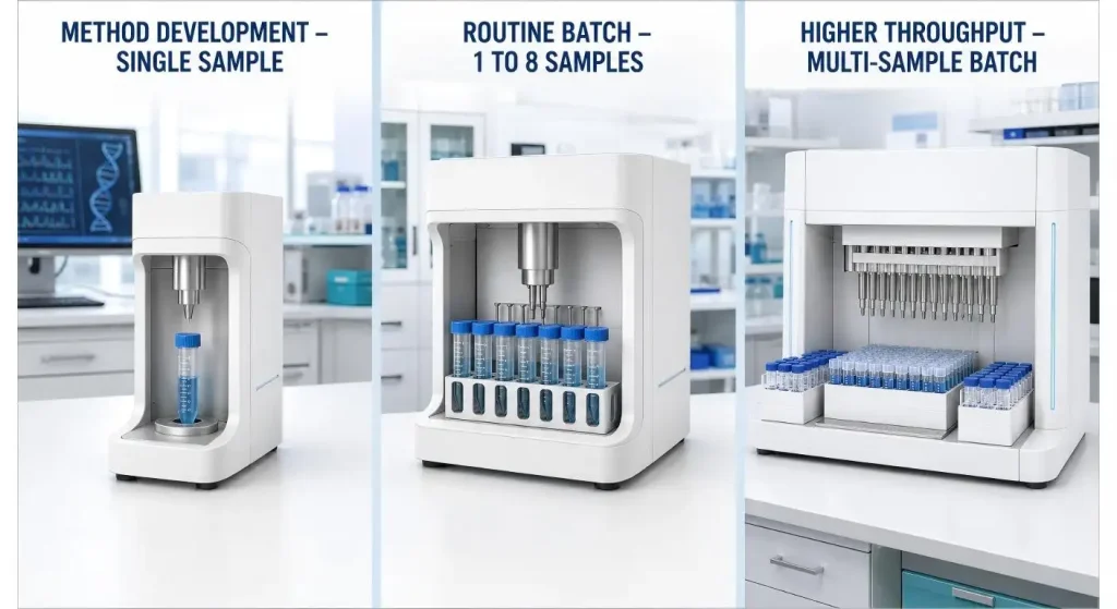
A single sample instrument can be appropriate for low-volume research or method development or for laboratories with fluctuating methods. Provides flexibility to the researcher in case the samples cannot be processed all at once.
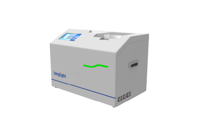
For laboratories which regularly process larger sample sets multi-position systems may be more suitable. The BoFU-800 multi-channel focused ultrasonicator is able to process 1–8 samples and provides individual and batch processing modes.
Don't consider only the number of tube positions as a measure of higher throughput. Other factors that buyers should also take into account are processing time, set up work, parameter management, sample traceability and if all positions can achieve the same fragmentation results.
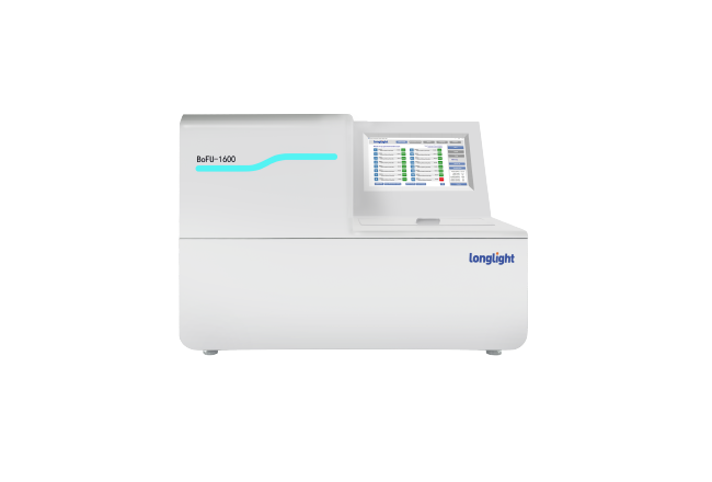
Another workflow is the BoFU-1600 multi-channel system, which is suitable for larger batch requirements.
Why are Tubes そして Sample Volume Important?
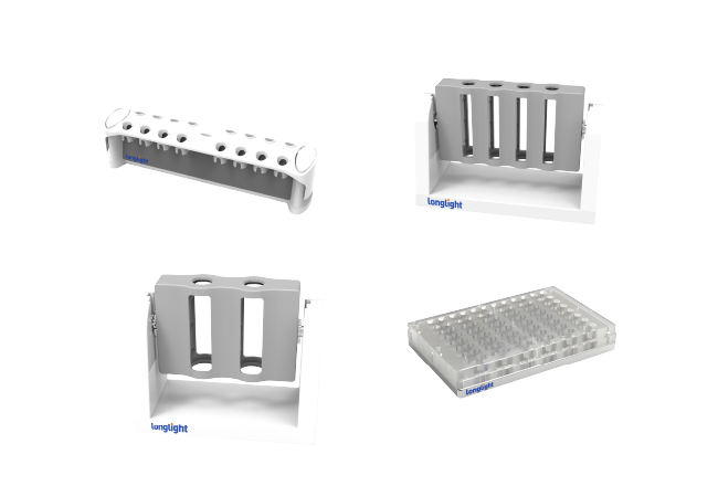
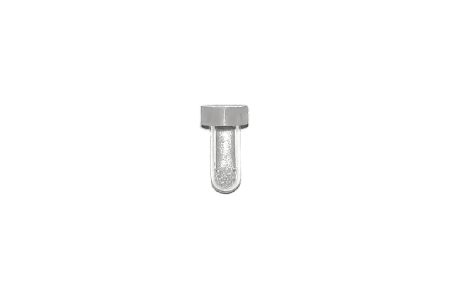
The sample tube is used as an acoustic system. The material that it is composed of, the thickness of the walls, the shape and the position in which it is located are all factors that determine how the ultrasonic energy is delivered to the liquid.
Instrument parameters may not be the same if an unvalidated tube is used, which will impact fragmentation performance. Closed tubes also minimise aerosol formation, cross contamination in sample processing.
The working volume should remain within the recommended volume for the tube selected. Fill volumes that are either too low or inconsistent may alter the acoustic focus and may have reduced repeatability of results.
How is a Fragmentation Method validated?
The representative sample material is the starting point to begin method validation. Use the suggested tube, volume, concentration and acoustic parameters for several replications.
| Record Item | Information to Save | Purpose |
| Sample identity | Type and extraction method | Tracks material differences |
| DNA input | Concentration and total mass | Supports method repeatability |
| Processing format | Tube type and sample volume | Controls acoustic transfer |
| Instrument program | Time, intensity and duty settings | Reproduces the method |
| 温度 | Start and process condition | Identifies heat-related variation |
| QC result | Size distribution and recovery yield | Confirms acceptance criteria |
Assess fragment size distribution, recovery yields and library performance downstream. However, the average fragment size is not sufficient – A high-molecular-weight tail may indicate under-fragmentation, whereas an excessive low-molecular-weight fraction may indicate over-processing or pre-existing DNA degradation.
Once a method is chosen set acceptable limits and document the instrument program. Routine controls are used by laboratories to alert them to changes in sample quality, consumables or instrument condition.
What is it Buyers should compare between Instruments?
Compare fragment ranges that can be achieved, sample volumes, tube formats, temperature-control method and number of positions for processing. Be mindful of protocol storage, record keeping, calibration and maintenance needs, too.
Request application information from suppliers from a sample that is similar to yours. Results of demonstrations should assess repeatability of tubes, repeatability of positions and repeatability of batches of runs, not just one successful run.
Availability of consumables and technical support are also crucial. An instrument that depends on difficult-to-source consumables can create workflow delays.Contact the Longlight contact team to match the BoFU configurations and consumables to the particular needs of the sample processing.
結論
The most useful application of an ultrasonic gene fragmenter is to make DNA or chromatin shearing a controllable, repeatable process! A focused ultrasonicator can make DNA or chromatin shearing a more controllable and reproducible process when energy delivery, temperature, tube format and sample conditions are properly standardized.
It is important to choose the right tool in regard to the actual samples in the lab and subsequent work or workflow. After the equipment has been installed, it is important to follow exact protocol to ensure that libraries are consistently good.
FAQ
Q1. Does an ultrasonic gene fragmenter cut specific DNA sequences or shear DNA in a largely sequence-independent manner?
No, the fragmentation that occurs with ultrasonic energy will be random mechanical fragmentation. Enzymes or programmable nuclease systems are needed to cut sequence specific.
Q2. Are the same settings to be used for all the DNA samples?
Not always. Changes in the behavior of fragmentation may occur as a function of the type of tubes used and the volume and concentration of DNA as well as the quality of the starting material; new sample conditions should be confirmed.
Q3. Can NGS be done with focused ultrasonication?
Yes. It was popularly used for controlled distribution of DNA fragment sizes for NGS library preparation. The target range should be the same as the library protocol selected.
Q4. What is a benefit of the closed sample tubes?
Closed tubes minimize direct contact with samples, and minimize aerosol formation and contamination. They also offer a controlled geometry in order to have a regular transfer of acoustic energy.
Q5. How should checks be made of the results of fragmentation?
Analyse the full size distribution by an appropriate method of electrophoresis or fragment analysis. These should also be checked for recovery yield and performance of the downstream library.
