

関連記事
明確で再現可能な結果を得るために適切なUVゲルイメージングシステムの選定
2026-09-16If bands are faint, background is high, or images are poorly exposed, even a successful electrophoresis run can be difficult to interpret.Imaging quality is a critical factor for the verification of results, recovery of the sample, reporting and for experiments with reproducible results for laboratories performing DNA, RNA or protein gels.
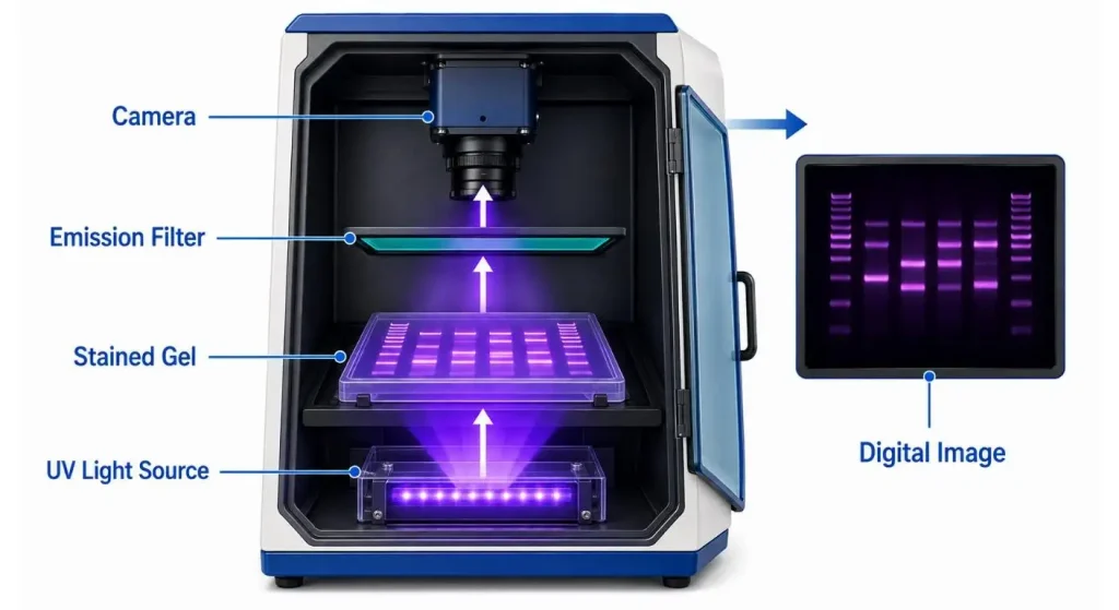
A UV Gel Imaging System is composed of the following components: controlled illumination, light-tight cabinet, optical filters, sensitive camera and analysis software. Thus, it is not enough to compare camera resolution when deciding which system to go for. The buyer has to take the whole imaging process into account, from stain excitation to the export of the data.
So What is a UV Gel Imaging System?
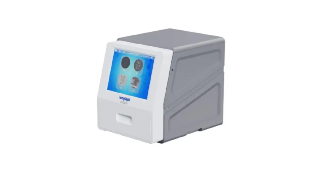
A UV gel imaging system is designed for the visualization and documentation of DNA, RNA, or protein bands after gel electrophoresis. By combining UV illumination, a high-sensitivity camera, optical filters, and image-analysis software, the system allows users to clearly capture gel images and analyze band position, brightness, and molecular size. It is commonly used in PCR verification, nucleic acid electrophoresis, restriction digestion analysis, and routine molecular biology experiments.
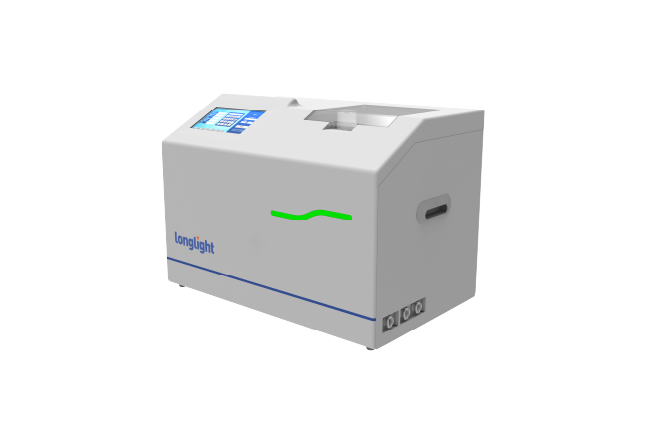
An enclosed imaging system will allow for controlled capture, analysis, labeling, storage and export, not possible with a simple UV viewing box. The Gel Imaging System (GI-200) from Longlight combines all of these features in one benchtop imaging system for everyday laboratory imaging.
Why Illumination and Stain Compatibility Matter are Important
A good UV source is not necessarily the best for making a good image. The excitation wavelength should correspond to the selected stain and there should be an optical filter which passes the emitted fluorescence. This mismatch may result in dim bands, high background or false differences between lanes.
302 nm UV is commonly used for many conventional nucleic-acid stains, while other wavelengths or blue light may be preferable depending on the dye and downstream application. It is important laboratories confirm the compatibility of the stain, and not use one wavelength for each assay. The Longlight guide to choosing a UV transilluminator informs you of the wavelength, illumination uniformity and viewing area and how they affect results.
Assess Camera as a Complete Imaging Chain
Megapixels are used to indicate image dimensions, but aren't performance indicators alone. The ability to record faint and strong bands in the same image is a function of the sensor's sensitivity and signal-to-noise ratio along with the quality of the lens and choice of filters, dynamic range, and uniformity of the illumination.
If routine PCR testing is performed, clear band separation might be adequate. For samples with low abundance and semi-quantitative comparisons, lower noise and greater dynamic range is required. Users should ask to see pictures taken by the user with their type of gel and stain rather than relying on a resolution sheet.
DNA protection during Imaging and Gel Excision
Nucleic acids may be damaged by UV exposure, particularly if the gel is still exposed to UV during the process of cutting the bands. This can decrease the efficiency of cloning or have an impact on sensitive downstream applications. Therefore, exposures should be minimized.
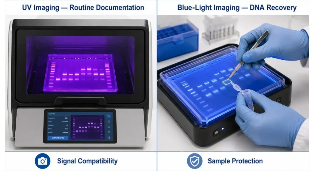
Lab flexibility is provided by a system which provides two modes: UV and blue-light. UV is suitable for well-known stains and for regular documentation, but Blue light can be preferable when DNA will be recovered from the gel, provided that a blue-light-compatible nucleic-acid stain is used.For further details see Longlight's high resolution gel imaging and UV-safe gel excision guide.
Make UV Safety a Purchasing Requirement
The eyes and skin can be damaged by the direct or reflected UV radiation. A closed imaging cabinet with UV-blocking panels and door interlocks can reduce accidental exposure to UV radiation.
Laboratory procedures are not a substitute for safety features. However, appropriate personal protective equipment, training, warning signs and documented operating practices are required of users. But a properly designed enclosure will make it easier to behave correctly and reduce the exposure during the routine imaging.
System matches the Laboratory ワークフロー
Laboratories should establish their desired outcome of the resultant image before making a purchase. Maybe the teaching lab needs to see the results quickly, or perhaps the regulated or research lab might want repeatable acquisition settings, user records, annotations and searchable files.
Benefits of the workflow include autoexposure, auto focus, ladder detection, image optimization, automatic file naming, data transfer and audit trails. Consider multi-light-source electrophoresis gel imaging for the multi-assay lab, which also can reduce the need for separate instruments.
| Evaluation Item | Basic Requirement | Question for Supplier |
| Sample coverage | Supports routine gel dimensions | What is the usable imaging area? |
| Light sources | Compatible with current stains | Are UV, blue, and white light available? |
| Weak-band capture | Low-noise, sensitive detection | Can you image our test gel? |
| ダイナミックレンジ | Avoids strong-band saturation | Can faint and strong bands be captured together? |
| 安全性 | Enclosure and UV protection | Is UV operation interlocked? |
| Software | Capture, annotation, and export | Can validated methods be saved? |
| Traceability | Retains settings and user records | Are metadata and audit logs available? |
| Support | Training and maintenance options | What local service is provided? |
Try out with the real samples!
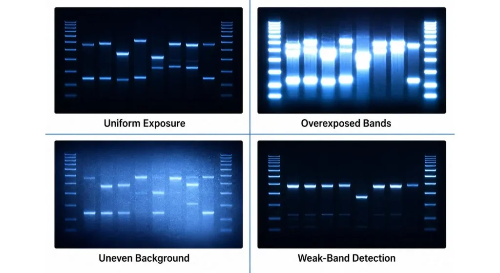
A demonstration should include not only a "laboratory" gel, but also simulate "normal" conditions. Test weak and strong bands side by side, compare the same sample bands on different parts of the stage and compare the automatic setting to a validated manual setting.
See if the system detects faint bands without saturating strong bands. Consider background uniformity, focusing consistency, annotation accuracy, export speed and ease of cleaning as well. These tests should show the real-world performance better than spec-sheet cameras.
Standardise Imaging post installation
Even the most competent system will vary from one user to another if the users alter the stains, exposures, filters and any other file handling without documenting it. Laboratories should develop imaging techniques that are specific to each application and save validated settings, if provided by the software.
The following routine checks are recommended: Imaging Surface, Illumination Output, Focus, Filters, Storage Capacity, and Safety Interlock. It is also useful to record the dates of maintenance as this can help to differentiate instrument drift from staining or sample preparation changes.
If you already have a system, what are the signs it's time to get a new one?
Replacement can be warranted if faint bands are consistently missed, there is uneven illumination, exposure settings cannot be replicated, and/or image files are missing metadata to facilitate review. The ongoing maintenance needs and the fact that older platforms may not be as stain resistant as newer ones can also add up to the total cost of having an older platform.
Repeat experiments, operator time, safety limitations and data-management needs should be used to make the decision – NOT just equipment age. Laboratories considering an upgrade can call Longlight to discuss their samples, stains, gel formats and documentation requirements.
結論
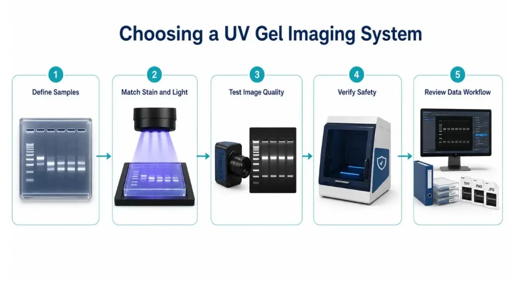
The highest resolution camera isn't necessarily the best UV Gel Imaging System. It is the system that matches up light with stain chemistry, avoids band saturation, safeguards the user and the samples and leaves consistent records.
Laboratories can use a sample-based demonstration or a specification review that focuses on workflow to choose a platform that will provide reliable results between operators and experiments.
よくある質問
Q1. What's the purpose of a UV Gel Imaging System?
It captures stained DNA, RNA or protein gels after electrophoresis.The images created are then analysed, documented, compared and exported.
Q2. Does it make sense to use UV imaging for DNA to be recovered?
It could be used, but there should be an effort to minimize exposure as UV can damage DNA. For gel excisions and subsequent cloning it is often preferable to image in blue light using a compatible stain.
Q3. Why is it that in the captured image the gel bands are faint?
This could be due to low sample concentration, stain–light mismatch, inappropriate filter, improper exposure, old lights, or high background light. The entire optical set-up should be verified prior to repeating the experiment.
Q4. The more megapixels a camera has, the better the picture is right?
Not at all, there are several other factors such as sensitivity, noise, dynamic range, quality of lens/filters used, and uniformity of illumination. Check real-life photos instead of megapixels.
Q5. What is the recommended frequency for imaging checks of an imaging system?
Routine checks should include imaging-surface cleanliness, illumination uniformity, focus, filters, safety interlocks and image consistency.
