

関連記事
DNA抽出用磁気ビーズセパレーター:実践的な選択ガイド
2026-09-16Most of the focus on DNA extraction is on magnetic bead chemistry. However, the separator that contains the tubes can also impact recovery, purity, and operator consistency.
Capture time and capture-zone consistency are important performance factors for a magnetic bead separator that is required to immobilize beads bound to the DNA in an efficient manner without compromising the beads' predictable immobilization position during aspiration. In the case of a weak, irregular and/or poorly visible capture zone, beads may be lost with the supernatant or may be suspended in wash buffer.
How does Magnetic Bead DNA Extraction work?
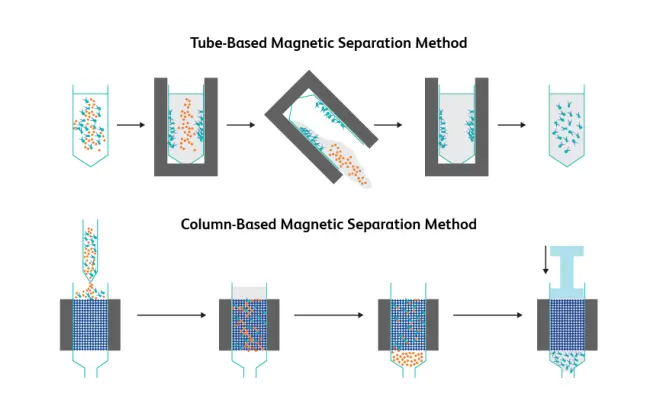
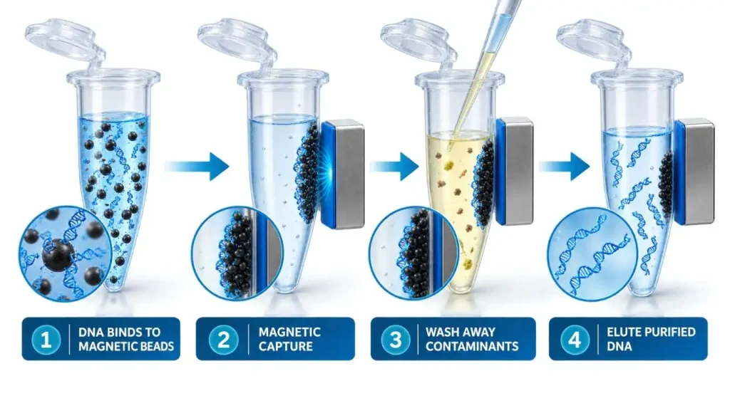
Magnetic bead extraction involves the use of paramagnetic particles that have surfaces which bind nucleic acids under well-defined chemical conditions. After lysis, the appropriate binding conditions promote adsorption of nucleic acids onto the functionalized magnetic beads.
The magnetic field exerts a force on the beads bringing them to the bottom of or towards the wall of the sample vessel. The supernatant can then be removed by pipetting without centrifugation.Before the DNA is eluted into the elution buffer, salts, proteins and inhibitors are washed out of it by wash buffers.
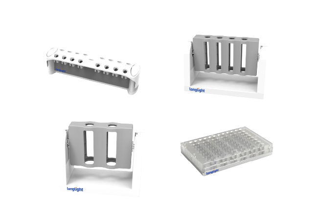
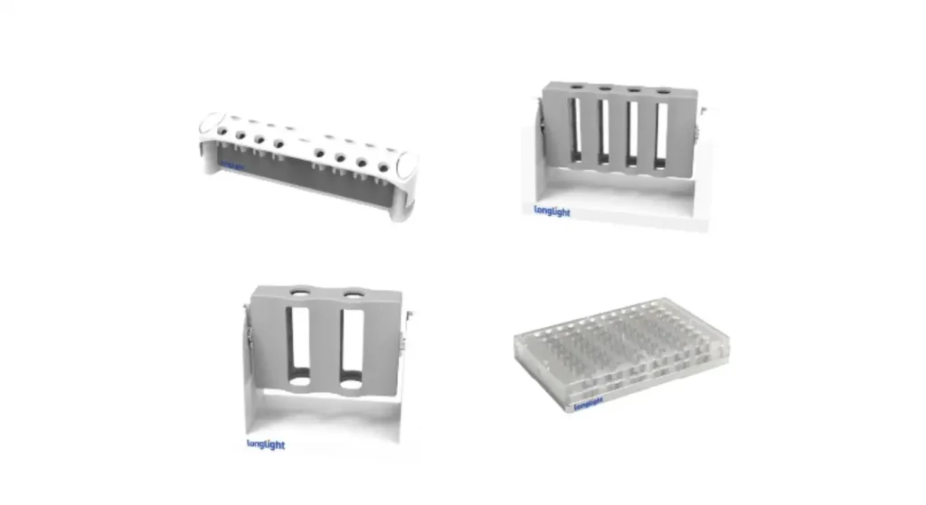
The separator is not involved in the chemical binding. The purpose of this is to reliably immobilize the beads during steps of separation and liquid removal. The MSG磁気ラック from Longlight is a permanent magnet based tube separation device that separates nucleic acids, proteins and cells.
Why is Magnetic-Field Design important?
The performance of a separator cannot be determined by magnet strength only. The distance between the magnet and sample, magnetic-field gradient, tube geometry and bead properties all affect capture behavior.
A good field will concentrate beads to form a small, easy-to-see area. This will facilitate removal of the supernatant without disturbing the pellet while placing the pipette tips.
A too aggressive or uneven capture pattern can result in compact bead clumps which are not easy to resuspend. A poor setup may allow beads to be hanging for too long. The ideal aim is to separate quickly, thoroughly and repeatably without complicating the process of subsequent mixing.
What Are Some Places Where DNA Can Be Lost?
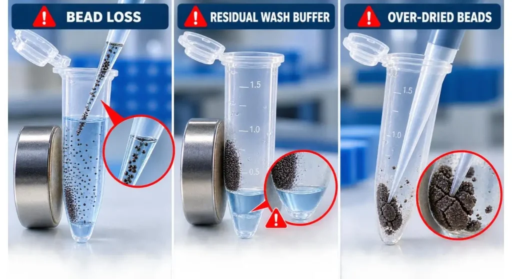
The most frequent loss of beads is when the supernatant is removed and washed. If the pellet is not visible, the pipette tip may displace it and/or suck it up.
The problem with the remaining liquid is different. Incomplete removal of the wash buffer can lead to carryover of ethanol, salts or other components, which may affect PCR, ligation or library-preparation reactions.
It is also important to not overdry the product. Over-drying the beads after the final wash can make them difficult to resuspend and may reduce elution efficiency.The clear capture zone guides the operator to the correct removal, and the next step in the right time.
What's the best match-up for Tube Format and Volume?
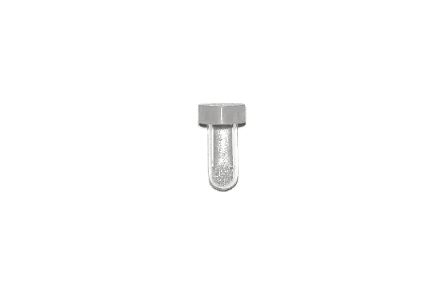
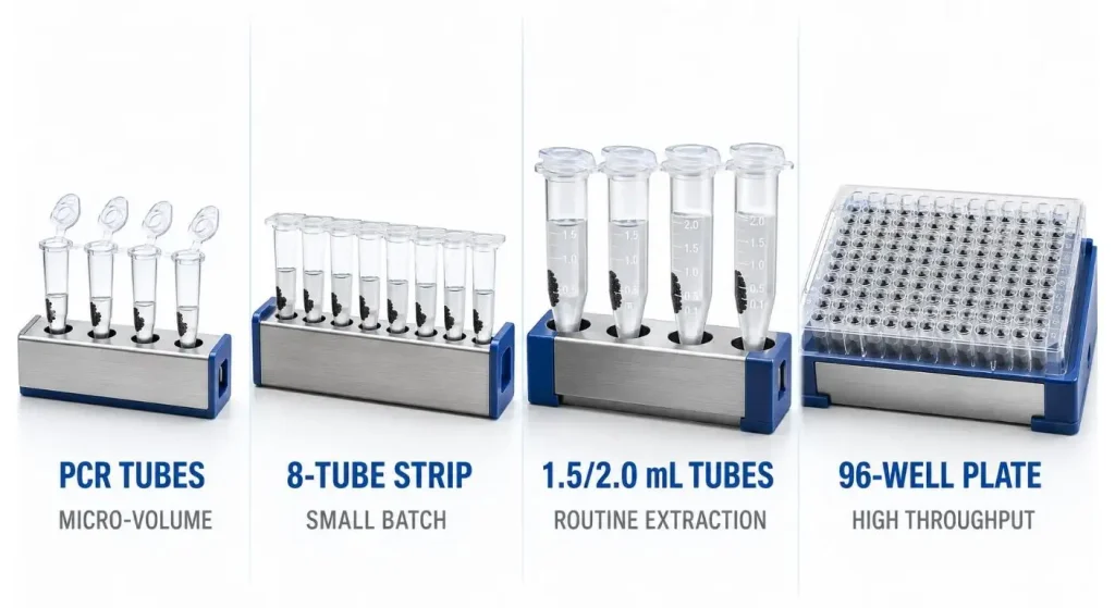
The separator should contain the actual vessels used in the protocol. These are typically in the form of PCR tubes, 8-tube strips, 96-well plates, 1.5 mL or 2.0 mL microcentrifuge tubes and larger conical tubes.
The location of a tube within a rack might be too far away from the magnet for another format. Good alignment is important even when the tube is physically able to fit as poor alignment can cause more separation time or an inconvenient location of the pellets.
Volume of work is important, too. Longlight's MSG series includes a variety of small volume formats ranging from microliter samples up to 2 mL in a variety of PCR vessels and standard microcentrifuge tubes.
What is the そうだ amount of throughput to have?
A research lab that is working with a handful of samples might want a small rack that is highly visible and has tubes that are easily accessed. The format must fit the diagnostic or sequencing laboratory and the size of the batch and the multichannel pipetting protocol.
Not using all positions is not necessarily an issue, but when an eight-tube protocol is used, differences in timing can occur between samples when applied to individual handling. Predictable bead capture across all positions is important for higher throughput, where consistent tube spacing is important as well.
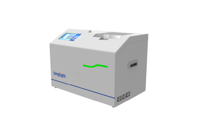
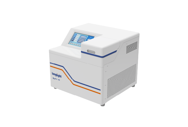
Longlight’s NGS sample-preparation portfolio covers magnetic separation, DNA quantification, DNA fragmentation, and electrophoresis, providing a more integrated workflow for sample preparation.
Washing – どう is it to be done?
After beads have congregated, put the pipette tip on the other side from the bead. Allow the liquid to be removed slowly enough so as to not pull any beads off the magnet.
Add the specified wash buffer following standardised protocol. Depending on the workflow, washing can be performed with the tube on the rack or the beads need to be completely resuspended between washing steps.
The method should indicate if the beads should be completely dispersed during each step. Contaminants may be trapped in clumps if the resuspension is not complete, and may cause greater variation in sample handling if mixing is overly aggressive.
How is DNA eluted off of the beads?
Once final washings and drying (controlled) are finished, remove vessel from the magnetic rack. Add indicated elution buffer and thoroughly resuspend beads.
The recovery of DNA may be affected by the temperature, elution volume, mixing and incubation time. When elution has finished, return the tube to the magnetic rack and wait until the beads are fully recaptured and the supernatant is clear before transferring the supernatant containing the DNA.
The elution position of the pellet is of particular importance if using elution of low volume. A compact pellet, which is farther out from the aspiration zone, results in more eluate recovered, but without the transfer of beads.
How can Separator Performance be measured?
Repeat extractions with the same sample, bead chemistry and person doing the extraction. Note time required for separation, visibility of pellets, ease of removing supernatant and ease of resuspension of beads.
Check the amount and quality of the DNA, and also test the application you intend to use the DNA for. Even if a sample has acceptable absorbance ratios, the sample may still contain a sufficient amount of inhibitor to impact PCR or sequencing-library preparation.
For multi-tube and plate, inter-position consistency is important. Test more than one test location – comparing results near the center and at the edges.
When Is a Larger Biomagnetic Separator Required?
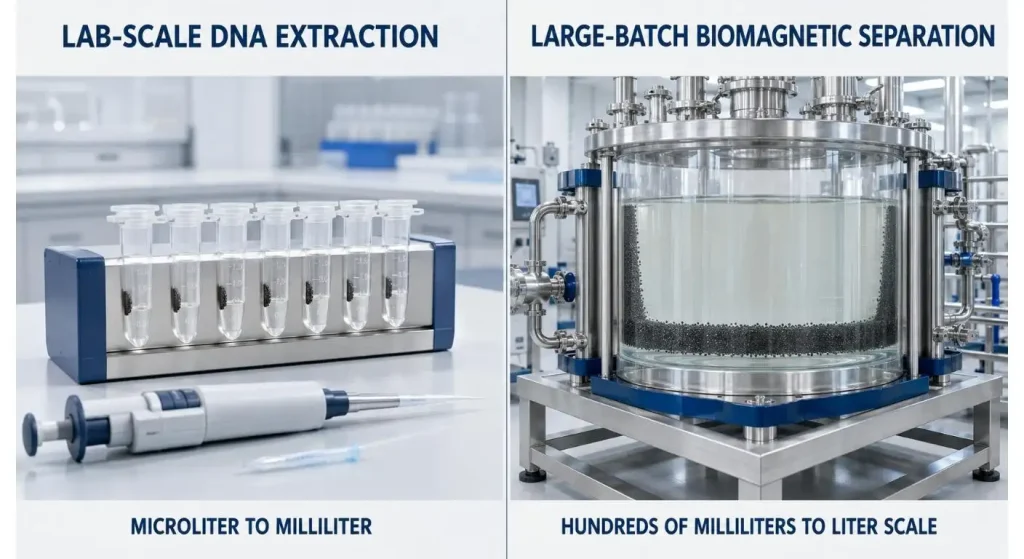
Tube racks can be used for everyday microliter to milliliter applications. Separation of hundreds of milliliters or a few liters of reagents, bioprocessing or manufacturing may be required.
| Observation | What to Record | Possible Warning Sign |
| Clearing time | Time until supernatant is clear | Beads remain suspended |
| Capture zone | Pellet location and shape | Diffuse or split pellet |
| Aspiration | Ease of removing supernatant | Beads enter pipette tip |
| Resuspension | Time and mixing needed | Persistent bead clumps |
| Final eluate | Volume recovered | Large volume loss |
| Position consistency | Yield across rack positions | Edge-to-center variation |
It's not as if a larger magnet will make a difference at that order of scale. Vessel diameter, mixing, bead travel distance, field uniformity and process monitoring are more significant.
Longlight's biomagnetic separators, model MSG-250/MSG-1000 are useful for scale-up for separation of nucleic-acids, protein purification and other batch processes.
Buyers Checklist.
Verify the following: tube types that are supported, working volume range, how many positions, and whether it is compatible with single-channel or multi-channel pipettes. Look at the location of the beads and if the pellet is still visible from the operator's normal work angle.
Request “separation data” in a bead and vessel format that is similar to your protocol. Furthermore, consider cleaning needs, chemical resistance, rack stability and ease of tube insertion and removal without abrupt movements.
A product family that includes several volumes can ease the training of laboratory personnel and method transfer for laboratories that use multiple extraction formats. Magnetic separation and others are Longlight's general laboratory workflow solutions.
結論
The accuracy of wash liquid removal and the amount of DNA-binding material which is left in the tube is directly influenced by the magnetic bead separator for DNA extraction. A visible pellet and a correct tube positioning help to achieve a higher recovery and better repeatability with the Uniform bead capture.
The separator must be considered in conjunction with the bead chemistry, sample volume and liquid-handling technique. Lab users can email their tube format and batch size and/or working volume requirements to the Longlight contact page for product matching.
FAQ
Q1. Does a magnetic separator bind DNA directly or does it bind to some other molecule that will bind to DNA?
No. The beads are coated with a surface chemistry that binds with DNA. The separator is able to retain these beads and draw off the liquid surrounding them.
Q2. Is there one magnetic rack for each type of tube?
Usually not. The working volume and tube geometry must be checked as the physical fit does not necessarily mean that the correct distance from the magnet is maintained.
Q3. What time should be allowed for magnetic separation?
It is dependent on the attributes of the beads, sample volume, viscosity, tube shape and magnet design. Perform the validated protocol, and wait until the supernatant is clear.
Q4。 Why are magnetic beads present in the final eluate?
May have disturbed the pellet during aspiration or too short a clearing time. Carefully put tube back in the magnet and transfer the supernatant again.
Q5. Is it possible to extract RNA using magnetic separators?
Yes with a suitable RNA binding bead chemistry and extraction procedure. Still, RNase-control must be performed.
