関連記事
UVトランスイルミネーター:ジェル可視化に適したシステムの選択と使用方法
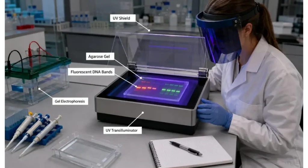
2026-07-30the results is problematic is a UV transilluminator. It reads DNA, RNA or protein bands in a simple way, by passing ultraviolet light through a stained gel. The operator can then examine the gel, take a picture of it, or cut out the desired band to work with it later.
Most issues relate to other than power switch. These are caused by incorrect wavelength, incompatible lighting and stain, too long exposure and lack of UV protection.
A good UV transilluminator workflow should provide clear bands, with only the minimum amount of exposure to the sample and the person using the instrument.

How a UV Transilluminator works
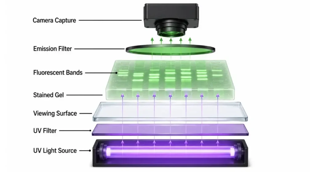
The gel is then electrophoresed, leaving behind molecular fragments which are usually not visible to the naked eye. A fluorescent stain that attaches to or interacts with the material to be stained. The stained gel is then transferred to the illuminated surface and the dye is excited by the ultra-violet light and gives off a fluorescence that is visible by the eye.
The dark background and bright bands allow you to approximate the size of fragments, compare the intensity of samples, check amplification products or find material for gel extraction.
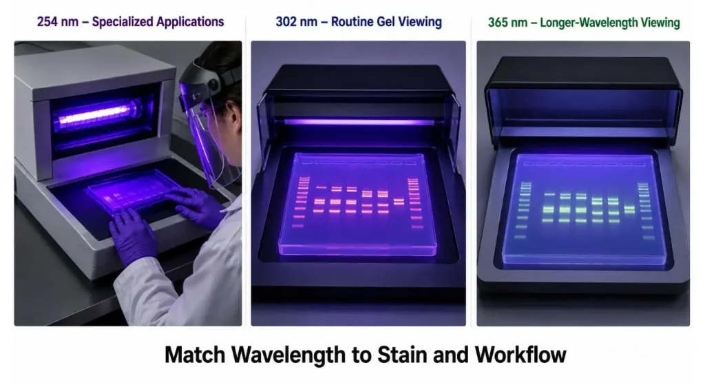
There are several UV transilluminators available at standard wavelengths of 254, 302 and 365 nm. Some offer a single wavelength, others offer two or three; which is best depends on the type of instrument and the wavelength desired. Gel viewing and electrophoresis applications utilize a wide range of UV near 302 nm.

Select the Wavelength for the actual Application
The intensity of the fluorescence, damage to the sample and stain compatibility are influenced by the selection of the wavelength.
A 302 nm source is often employed when viewing fluorescently stained DNA and RNA gels as it generally yields a high intensity signal and many known nucleic-acid stains can be used with this source. An easy solution for routine gel checks and documentation.
If the reduction of UV-induced DNA damage is more important than the strongest possible signal, it may be considered to use longwave UV, which is near 365 nm. Thermo Fisher advises to minimize exposure to DNA and suggests the use of a longer wavelength (around 360 nm) if DNA will be recovered from the gel.
The use of shortwave UV (near 254 nm) for routine viewing of DNA-gels should not be considered as automatic, although some lab transilluminators are available. It is suitable to use depending on the fluorescent material and the application in the laboratory.
The greatest wavelength is thus not the most effective option. It's the one that suits the stain and maintains the sample sufficiently for the following step.
| 選択因子 | なぜ重要なのか | 確認すべきこと |
| UV wavelength | Affects stain excitation and sample exposure | Stain compatibility |
| Viewing area | Determines supported gel size | Gel dimensions |
| Light uniformity | Influences band consistency | Full-surface illumination |
| Intensity control | Helps limit unnecessary exposure | Available settings |
| Protective shield | Reduces direct UV exposure | UV-blocking design |
| Imaging compatibility | Affects documentation quality | Camera and filter support |
| Lamp maintenance | Influences long-term signal stability | Replacement and monitoring |

Use a different colour of gel stain for a light source of a different colour
A transilluminator and gel stain should be considered as one optical system.
There are different types of dyes with differing characteristics of excitation and emission. A stain might have a high UV reflectivity, and low blue light reflectivity, and vice versa. The camera filter should also allow the emitted fluorescence to pass through and prevent the passage of unwanted excitation radiation.
Prior to a new assay:
- The wavelengths that excite the stain are lequel.The wavelengths that excite the stain are 'luequels'.
- The decision whether or not to use a photographic filter will be determined by the individual photographer.
- The attributes of the staining substance (DNA, RNA or protein)
- If the gel is to be cut or not, how will it be documented?
- If the light source is a new technology, is there a method for capturing it using the current imaging system?
For instance, SYBR Safe stained gels can be imaged using appropriate fluorescence equipment with either a UV or blue light filter - a filter used for ethidium-bromide imaging may not be suitable.
If an image is just too faint, it doesn't necessarily mean that there is too little DNA in the gel. The cause can be an inappropriate excitation wavelength, wrong camera filter, used out-of-date UV lamps or too much room light.
If the damage to the DNA will be recovered, limit the exposure to UV rays
Nucleic acids are susceptible to damage by UV light. This is significant if a researcher wishes to extract a DNA band from the gel and utilize the remaining DNA in a downstream application such as cloning, ligation, sequencing, or other applications.
Long exposure can lead to the nicking of the DNA as well as other damages, which can affect the quality of recovered DNA. Exposure to UV for extended periods may result in damage to DNA which could affect downstream applications such as subcloning, Thermo Fisher points out.
In the event that UV visualization is required:
- Set up cutting tools and collection tube prior to turning on the light.
- Set the lowest setting which will produce a clear band.
- Exposure should be kept to a minimum.
- If possible, think of a longer wavelength for the UV light.
- Do not repeatedly view the same gel.
- Make note of the chosen band and turn off the source.
A blue-light transilluminator that is compatible for cloning may be more suitable for particularly sensitive cloning work. Colors can be excited by blue light, and less damage has been found to be done to the DNA by blue light.
Check Illumination Uniformity
The transilluminator should provide uniform illumination of the area to be viewed.
If one side of the surface is more intense than the other, identical bands will be the varying intensities depending on their location on the gel. This is particularly relevant when lanes are compared in the same laboratory and/or when an image is employed for semi-quantitative analysis.
The uniformity is influenced by the condition of the lamps, the arrangement of the lamps, filters, cleanliness of the surfaces and the optical design of the instrument. Uniform illumination is important in the context of UVP for achieving repeatability of results in various bands and lanes.
Always examine the background to see if it is even throughout the entire gel, before depending on the band intensity. Transferring the same gel to a different part of the surface may help to develop a problem of illumination.

Use the correct viewing area
The area the surface is lit should be sufficient to cover the gel but not take up any extra bench space.
A little cell is great for standard mini-gels. Bigger platforms will be better for large format gels, or multiple gels side by side. There are commercial systems available with different illuminated areas, typically of about 20 x 20 cm. and greater.
All the gel should be flat on the surface. Reflections and blurring of the bands are possible due to wrinkles in the protective film, too much buffer or an uneven tray.
Ensure viewing surface is clean; only use materials and cleaning agents that are compatible with the UV filter. Images may be affected by scratches, stains or dried buffer deposits.

Make sure to protect the user from UV exposure
A UV Transilluminator is NOT a standard viewing lamp, but a light source with high intensity which is designed for use in the laboratory.
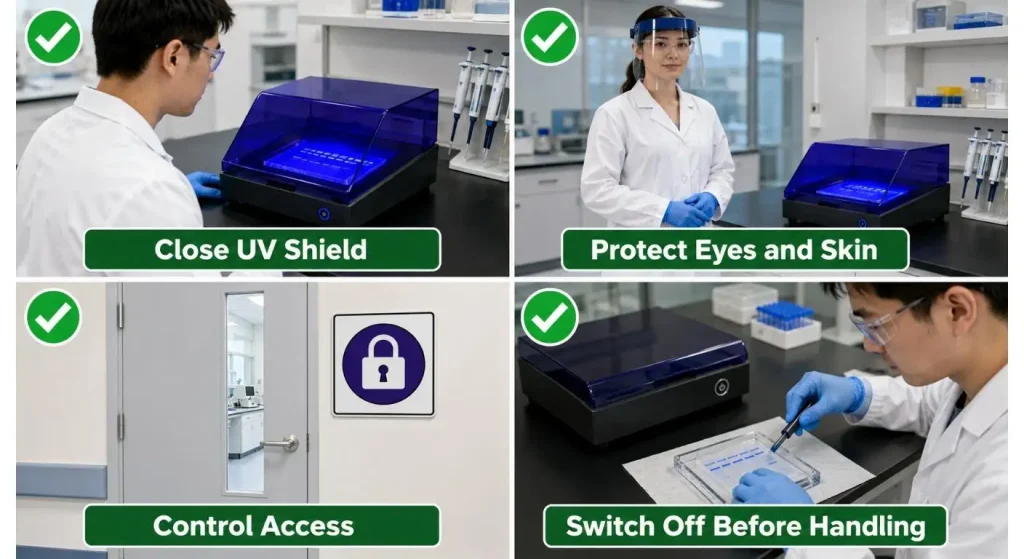
Either direct or indirect exposure to UV radiation can lead to eye and skin damage. The laboratory safety guidelines of Stanford list the following precautions for using a transilluminator: keep access to the transilluminator under control when it is in use, keep reflective surfaces away from transilluminator, and post signage indicating that skin and eyes should be protected.
Always utilize the protective top, shield or enclosed imaging cabinet provided with the instrument. Laboratory practices that require working with the gel may necessitate covering the face, wearing gloves, a fastened laboratory coat and covering the skin with UV blockers.
Don't presume that normal safety glasses block the UV wavelength. Wear appropriate protective equipment that has been designated for the instrument and adhere to the lab's radiation-safety rules.
Better exposure control is achieved with an enclosed gel-documentation system with safety interlock as opposed to working over an open light box. Some systems do not allow the imaging door to be opened while operating in the UV mode.
Educators should establish a consistent viewing workflow
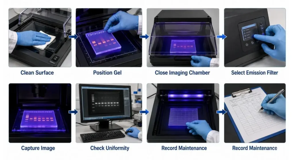
There's a simple track that can be used to minimize unnecessary variability:
- Electrophoresis and Staining 1.Electrophoresis and 1 staining.
- Ensure that the stain is the correct wavelength.
- Make sure the outside of the gel tray is clean and dry.
- Choose the wavelength and intensity of UV required.
- Lay the gel on the viewing area in the center.
- Shut the protective shield/imaging enclosure.
- Employ the shortest exposure time possible to produce visible bands.
- Take the photo with the appropriate emission filter.
- Turn off the UV light when moving the gel.
- Wipe down surface after use.
If performing several experiments, the stain concentration should be the same, as should imaging filter, exposure time, intensity setting and camera conditions.
Plug the Transilluminator into the Imaging Workflow
If the user does not require to inspect a gel, then a benchtop transilluminator can be used. For more demanding applications a full gel-documentation system including a camera, dark enclosure, filters and image-analysis software is needed.
Integrated systems can be used for image capture, band measurement, documentation and regular gel analysis. Bio-Rad characterizes gel-documentation platforms, which feature controlled illumination and automatic image acquisition and analysis software.
The decision will depend on what the lab is going to do with the image. In a research laboratory, the teaching laboratory might need a clear visual confirmation, a research laboratory might need traceable image files, consistent exposure, and quantitative band analysis.
Never remove the Light Source
Even if UV lamps look like they are on, they begin to lose their output over time. With reduced intensity, the operator will then increase the exposure time, which could lead to less consistency, and more sample exposure.
Follow manufacturer's lamp maintenance guidelines. A UV radiometer would be a more objective measure than visual estimation and UVP has determined that irradiance measurement is an appropriate method for monitoring for the useful life of a UV source.
Change lamps as a set if recommended, thoroughly clean the viewing filter and note dates of maintenance. Fluorescent UV lamps can also contain mercury and should be dealt with and disposed of as a hazardous waste per local requirements.
結論
When using a UV transilluminator for gel visualization, it can do it quickly and clearly, but it cannot only be bright in ultraviolet light to obtain good results.
The wavelength should be appropriate for the stain and downstream uses. The illumination should be as uniform as possible, the exposure should be as short as possible, the camera filter should be the same as the emitted fluorescence, and the user should be protected from UV exposure from the light source.
The right instrument is the one that will accommodate the size of the gel, a staining method, the imaging requirements, the workflow for recovering the samples and safety controls for the laboratory environment.
よくある質問
Q1. A UV Transilluminator is used for what?
It is primarily used to visualize bands of stained fluorescently labelled DNA, RNA or protein in electrophoresis gels. It also can be incorporated into a gel-documentation system.
Q2. Which of the following wavelengths is commonly used for DNA Gels?
A common use of midrange UV (near 302 nm) is for routine viewing of the gels. The final selection should be similar to the stain, as well as the application down stream.
Q3. Does UV Light Mutate DNA?
Yes. Long exposure may cause DNA damage and decrease the cloneability of the DNA and downstream applications.
Q4. Which is more effective: Blue Light or UV Light?
If UV damage to the user and/or DNA damage is a concern, use of blue light is recommended for compatible stains. Even for some known stains, UV might give better excitation.
Q5. Why is there a weakness in the Gel Bands?
This is likely to be due to low sample concentration, an inappropriate stain, an inappropriate wavelength or filter, ageing lamps, too much background light, or poor staining.